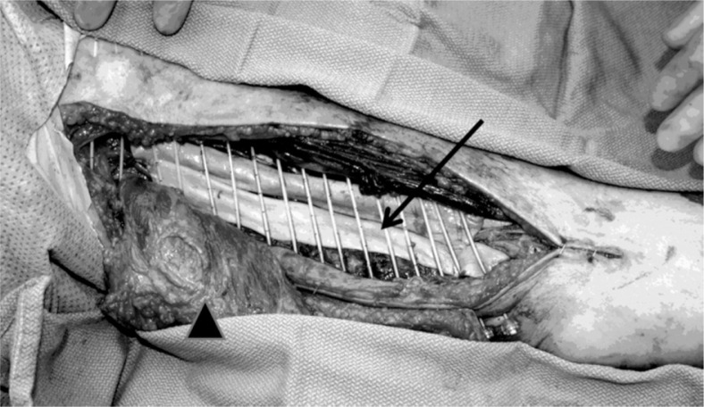
Fig. 1.
Intraoperative placement of catheters in the patient after a wide resection and latissimus dorsi myocutaneous rotational flap reconstruction. “→” identifies the Alloderm graft placed over neurovascular bundle. “▲” identifies the reconstructed flap which was subsequently rotated into the defect

