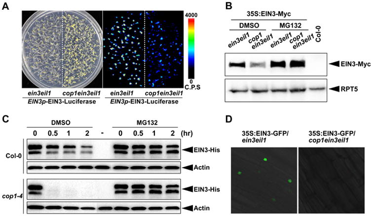
Figure 3. COP1 stabilizes EIN3 protein.

(A) White light (Left) and bioluminescence (Right) images of 4-day old etiolated seedlings of EIN3p-EIN3-Luciferase in ein3eil1 and cop1ein3eil1 backgrounds. The color-coded bar indicates the intensity of lucifersase activity. C.P.S stands for Counts Per Second.
(B) Western blot analysis of EIN3 protein levels. Seedlings over-expressing EIN3-Myc in ein3eil1 and cop1ein3eil1 backgrounds were grown on 1/2MS medium in the dark for 4 days without (DMSO) or with MG132 pre-treatment for 12 h before harvesting. Col-0 was used as a negative control. RPT5 was used as a loading control.
(C) Cell-free degradation of recombinant EIN3-His proteins in 4-day old etiolated Col-0 (top) and cop1-4 (bottom) seedlings. Equal amount of EIN3-His proteins were added into the cell extracts and incubated for the indicated periods of time, and then analyzed by immunoblots. “-” stands for the no EIN3-His protein control. Actin was used as a loading control.
(D) Fluorescence microscopic analysis of the EIN3-GFP protein levels. Seedlings over-expressing EIN3-GFP in ein3eil1 and cop1ein3eil1 backgrounds were grown on 1/2MS medium for 4 days in the dark.
