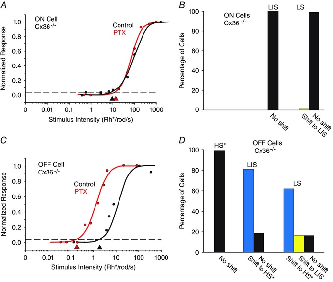
Figure 9. Ablation of certain rod pathways in the Cx36−/− mouse retina indicates that the three pathways carry rod signals with different sensitivities .

A, the intensity–response function of an LS ON RGC in the Cx36−/− mouse retina is unaffected by application of PTX. This is likely to be attributable to the fact that the primary and secondary rod pathways are dysfunctional in the Cx36−/− mouse. B, histogram showing that PTX did not increase the sensitivity of ON LS cells in the Cx36−/− mouse retina, a clear contrast from the increased sensitivity seen in the wild‐type (WT). C, application of PTX shifted the sensitivity of an OFF LS cell by ∼1 log unit to the LIS range. D, summary of the effects of PTX on OFF RGCs in the Cx36−/− mouse retina. Most cells showed a shift in sensitivity to the range of presumed HS cells (*) that show reduced sensitivity in the Cx36−/− mouse. Although PTX increased the sensitivity of most LIS and LS cells, it did not change the sensitivity of presumed HS cells (*) in the knockout. [Colour figure can be viewed at wileyonlinelibrary.com]
