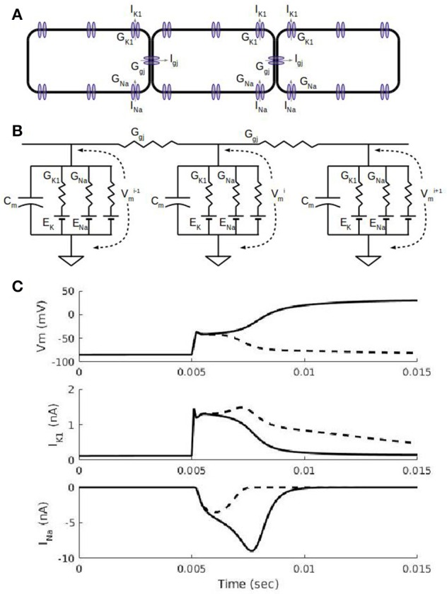
Figure 1.

Action potential propagation in a cardiac fiber. (A) Schematic view of three cells in a fiber. In addition to a number of sodium, potassium, and calcium currents in the cell membrane, each cell has non-selective gap junction channels that allow currents to flow down the fiber. The gap junction current, Igj, allows a depolarized cell to drive electrical activity in a neighboring cell. (B). Equivalent circuit representation of the three cells in (A). Each cell has a capacitance associated with the cell membrane and ionic currents represented by a conductance and a battery for the Nernst potential for the corresponding ion. The inward rectifier potassium current, IK1, current has a non-linear conductance (see Methods) with a maximal conductance, GK1, and its reversal potential is the potassium Nernst potential, EK. The fast sodium current, INa, has a time-dependent conductance with the maximal conductance, GNa, and its reversal potential is the sodium Nernst potential, ENa. (C) Simulations showing cell membrane potential, Vm, and ion channel currents, IK1 and INa, for a subthreshold stimulus (dashed curves) and suprathreshold stimulus (solid curves). Results shown are for the 5th cell in a fiber of 100 cells with 0.2 ms-duration current stimuli administered at the 5 ms mark to the first 4 cells. A subthreshold stimulus brings Vm to about −60 mV after which Vm declines (dashed curve) back to its resting membrane potential of about −90 mV; the IK1 waveform due to a subthreshold stimulus is larger than the suprathreshold case (solid curve) because the subthreshold Vms are more negative and thus less inward rectified. INa is partially activated (dashed curve) for a subthreshold stimulus and fully activated (solid curve) for the suprathreshold stimulus; the fully activated INa is what triggers the full depolarization of Vm to about +20 Mv.
