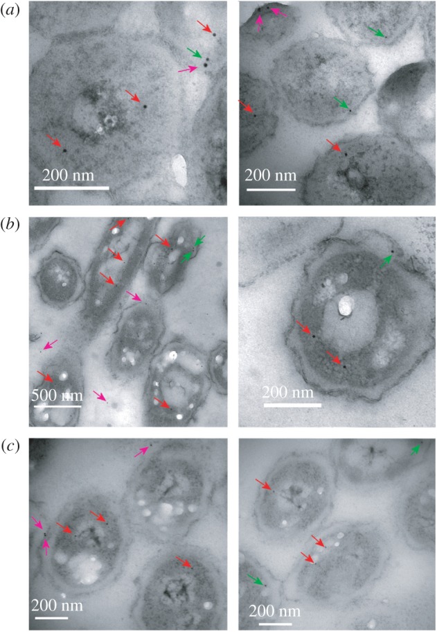
Figure 4.

Typical transmission electron microscopy images of E. coli cells treated with nanogold-conjugated FLAG antibody to reveal locations of PykF-FLAG. (a) 0 min, (b) 2 min and (c) 30 min. Arrows mark nanogold particles judged as cytoplasmic (red), inner membrane (green) and outside the inner membrane (purple).
