
Figure 1. THG sectioning image of a whole blood smear at 1-hour post blood sampling.
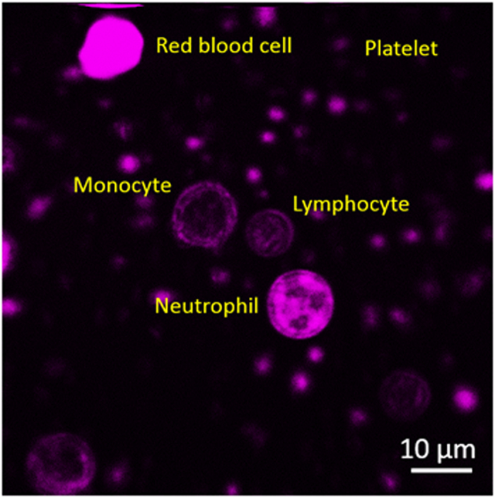
Different types of leukocytes could be distinguished by their size, THG intensity, and nuclear morphology. Field of view: 73 × 73 μm.
Official websites use .gov
A
.gov website belongs to an official
government organization in the United States.
Secure .gov websites use HTTPS
A lock (
) or https:// means you've safely
connected to the .gov website. Share sensitive
information only on official, secure websites.
Different types of leukocytes could be distinguished by their size, THG intensity, and nuclear morphology. Field of view: 73 × 73 μm.