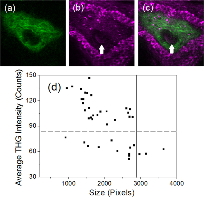
Figure 6.
The (a) SHG, (b) THG and (c) combined images of a capillary (indicated by a white arrow) in the dermal papilla region beneath the human skin. The papilla is surrounded by basal cells with strong THG contrast. (d) The scatter plot of the average THG intensity and size of WBCs’ cross-sectional images captured in the human capillary (See Fig. S9).

