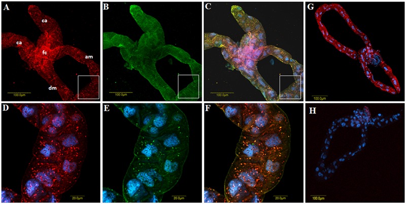
FIGURE 5.

Co-immunolocalization of TYLCV with first antibody against the virus CP and detected with secondary antibody conjugated to Cy3 (A, red), and CypB reacted with polyclonal anti-CypB first antibody and detected with secondary antibody conjugated to Cy2 anti-rabbit secondary antibody (B, green) in B. tabaci midguts dissected from viruliferous adults. (C) Shows the overlay of (A and B) and the yellow spots show the colocalization. (D–F) Are zoom in of the portions shown in the insets that appear in (A–C), respectively. (G) Is a control gut in which the whole co-immunolocalization procedure was performed without adding the anti-CypB primary antibody, and (H) is a control gut in which only secondary antibodies were used. ca, cecae; fc, filter chamber; am, ascending midgut; dm, descending midgut.
