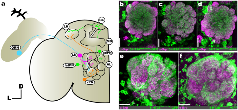
Figure 1. Consistent neuron labeling from different coding-intronic insertions of 5-HTRs in the antennal lobe of Drosophila melanogaster.
(a) Olfactory receptor neurons (ORNs; cyan) housed within the antennae and maxillary palps (not depicted here) send axons to a single glomerulus in the antennal lobe (AL). Within a glomerulus, ORNs synapse on projection neurons (PNs; green and orange) and local interneurons (LNs; magenta). LNs interconnect glomeruli and synapse on ORNs, PNs, and other LNs. A given PN is classified as an anterodorsal PN (adPN; green), lateral PN (latPN; green), or ventral PN (vPN; orange) based on its cell body position. PN axons project to the mushroom body (MB) calyx (Ca) and lateral horn (LH). Ellipses indicate neuron type, while circles indicate specific brain regions. (b–d) T2A-GAL4 conversion of three separate MiMIC insertions (4464, 1140, and 1468, respectively) in the 5-HT1A locus reveals consistent labeling of LNs and vPNs. (e and f) T2A-GAL4 conversion of two separate MiMIC insertions (6500 and 5208, respectively) in the 5-HT2B locus consistently labels ORNs. Neuropil in (b–f) are delineated by α-Bruchpilot (α-Brp; magenta) labeling. All scale bars = 20 um.

