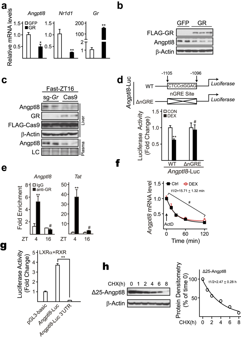
Figure 5. Identification of nGRE element in Angptl8 promoter region.
Cultured mouse primary hepatocytes were infected with Ad-GFP and Ad- GR virus, respectively. The infected cells were cultured overnight (O/N) in serum-free M199 and then treated with DEX (10 nM) for 4 hours. Angptl8 mRNA levels (a), and Angptl8 protein amounts (b) were analyzed by qPCR and immunoblot, respectively. (c) Knockout of Gr ameliorates Angptl8 reduction caused by fasting. Cas9 alone or sg-Gr-Cas9 adenoviruses were delivered into livers of adult C57BL/6 mice by tail-vein injection on Day1, and then animals were fasted at Day15-ZT12, euthanized at Day15-ZT16. Liver and plasma samples were harvested and immunoblot assay was performed to detect Angptl8 protein levels. (d) The nGRE site is necessary for Angptl8 to response to DEX treatment. Reporter plasmid (Angptl8-WT-Luc or Angptl8-ΔnGRE-Luc, up) and RSV-β-gal plasmid were co-transfected into HEK293T cells. The transfected cells were cultured O/N in serum-free DMEM and then treated with vehicle (PBS) or DEX (10 nM) for 4 hours. Luciferase activities were measured and normalized to β-gal activity (bottom). (e) ChIP analysis of the occupancy of GR on Angptl8 nGRE site in livers of ad libitum-fed mice euthanized at ZT4 and ZT16. Tat here serves as a positive control. (f) Half-life of Angptl8 mRNA. Cultured primary hepatocytes were treated with actinomycin D (ActD, 5 ug/uL) or ActD plus DEX (10 nM) for the indicated time points. Angptl8 mRNA levels were then analyzed by qPCR and Angptl8 mRNA half-life was calculated by using one phase decay equation. (g) 3′-UTR of Angptl8 takes responsibility for Angptl8 mRNA instability. HEK293T cells were transfected with Angptl8-Luc and Angptl8-Luc-3′UTR, respectively. The transfected cells were left overnight, and then lysed and assayed for firefly luciferase and β-gal activities. (h) HEK293T cells were transfected with Δ25- Angptl8 (Angptl8 without signal peptide) plasmids, then cultured O/N in serum-free DMEM and treated with cycloheximide (CHX, 10 ug/mL) for the indicated time points, after which immunoblot assay was performed (left) and Δ25- Angptl8 half-life was calculated by using one phase decay equation (right). Data are represented as mean ± s.e.m, n = 3, *p < 0.05, **p < 0.01, # no significant difference.

