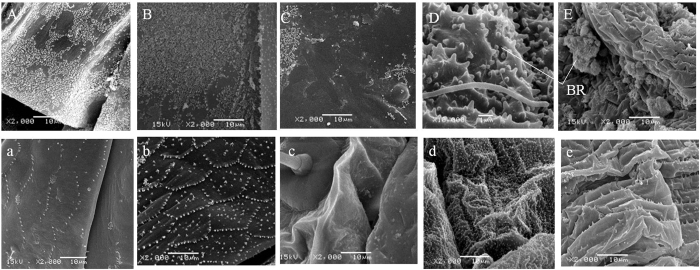
Figure 3. SEM images of the E. onukii abdomen.
(A–E) are images of abdomen cuticle with intact integuments, while (a–e) are images of abdomen cuticle with artificially bared integument. (A) and (a) represent adult dorsal abdomen; (B) and (b) represent the adult ventral abdomen; (C) and (c) represent nymph (1st to 5th) dorsal abdomen; (D) and (d) represent the ventral abdomen of younger nymph (1st and 2nd); (E) and (e) represent the ventral abdomen of older nymph (3rd to 5th); BR represent brochosomes. Scale bars: 1 μm for (D) and 10 μm for others. In (D) the size of brochosomes are similar to the micropapillae, and in (E) brochosmomes on the ventral abdomen of the nymph are clumped and have not been spread over its body.

