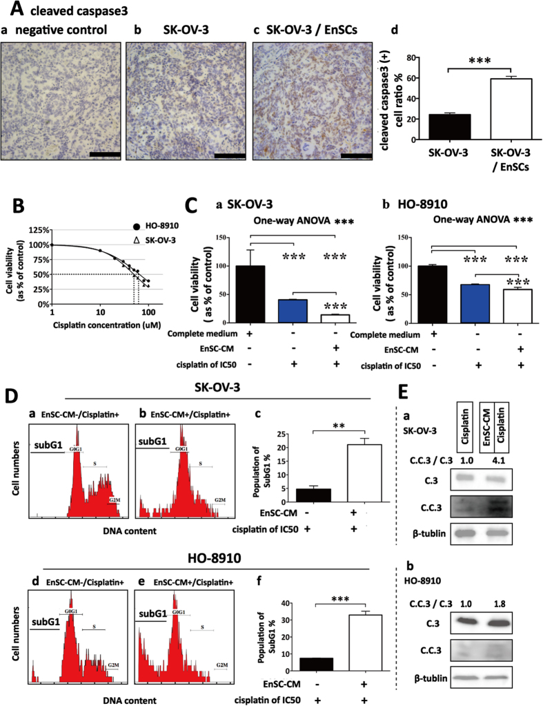
Figure 4. EnSCs promoted apoptosis in EOC cells in vivo and enhanced cisplatin-induced apoptosis in vitro.
(A) Apoptosis rate in tumor tissues was tested by IHC using antibody against cleaved caspase 3 (n = 5; Scale bar = 100 μm). Positively-stained cell ratios were measured and results were shown as averages of five randomly selected fields ± SEM. (B) The IC50 of cisplatin on SK-OV-3 cells and HO-8910 cells at the 48th hour was tested by CCK-8 assay (n = 3). (C) The effects of EnSC-CM on the cisplatin-induced cell death in EOC cells at the 48th hour were tested by using CCK-8 assay (n = 3; performed in triplicate). Ordinary one-way ANOVA was used for statistic analysis. (D) The effects of EnSC-CM on cisplatin-induced apoptosis in EOC cells were tested by subG1 assay using flow cytometry (n = 3; performed in triplicate). (E) Western blot analysis was performed on cell lysates harvested from EOC cells treated with cisplatin of IC50 with or without EnSC-CM for 48 hours by using antibody against both caspase 3 and cleaved caspase 3 (n = 3; performed in triplicate). The amount of protein loaded was normalized against β-tubulin. All data were shown as means ± SEM. **p-value < 0.01; ***p-value < 0.001.

