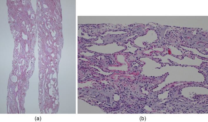
Figure 2.
Renal biopsy findings in case 1. (a) periodic acid-Schiff (PAS) stain; magnification 10×; (b) PAS stain; 200×. Light microscopy images show tubular dilatation and atrophy. Cystic tubules show irregularities and thinning of the tubular epithelial cells. The glomeruli show global sclerosis and arterial intimal thickening, consistent with the patient's age, without any other remarkable changes such as crescent formation.

