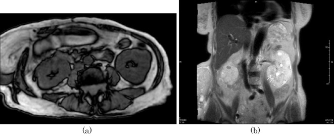
Figure 4.
Magnetic resonance imaging (MRI) of the abdomen: (a) axial section of the abdomen (T1-weighted image) and (b) coronal section of the abdomen (T2-weighted image) in case 2. No apparent microcysts are seen in the enlarged kidneys, including the coronal images, however, a closer observation shows multiple cysts along the corticomedullary boundary in the left kidney in the coronal section (arrows) (b).

