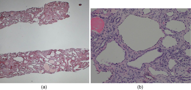
Figure 5.
Renal biopsy findings in case 2. (a) periodic acid-methenamine-silver (PAM) stain; original magnification 10×, (b) PAS stain original magnification 200×. Light microscopy images show tubular dilatation and atrophy. Cystic tubules show irregularities and thinning of the tubular epithelial cells. Of seven glomeruli, two are globally sclerotic, one is segmentally sclerotic, and two show glomerular cysts. The other glomeruli are intact.

