
Fig. 1.
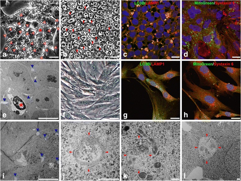
Autophagy machinery is widely operated in iPSCs. (a) Images of cells taken at day 15 of fibroblast reprogramming and large autophagic vacuoles indicated (red arrowheads). (b) Smaller autophagic vacuoles (red arrowheads) were observed in stable iPSC lines with daily change of culture medium. (c, d, g, h) Double immunofluorescence staining of iPSCs (c, d) and fibroblasts (g, h) was carried out with anti-LC3B for autophagy (c, g green) and anti-LAMP1 for lysosome (c, g red), Syntaxin 6 for Golgi membrane (d, h red) and MitoGreen (d, h green) for mitochondria, with counter-staining of DAPI (blue) for nuclei. Fluorescent images were acquired via confocal microscopy. Note that LC3B and LAMP1 are colocalized in iPSCs (c) but not in fibroblasts (g), whereas Syntaxin 6/MitoGreen are not colocalized as anticipated (d, h). (e, i–l) TEM images showed a dead nucleus (* a, e), lysosomal structures (blue arrowheads, e, i), and autophagic vacuoles (red arrowheads, j–l). Bar = 20 μm a–d and f–h; 10 μm e, i; 500 nm j–l