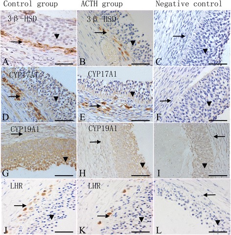
Fig. 4.

Localization of 3β-HSD, CYP17A1, CYP19A1, and LHR in sow ovaries Immunohistochemical staining of sow ovaries. Samples are counterstained with Mayer’s hematoxylin. Bars = 50 μm. The immunolocalization of 3β-HSD (a, b), CYP17A1 (d, e), CYP19A1 (G, H), and LHR (j, k) in the follicular walls of sows during the control and ACTH groups. The column on the right illustrates the immunohistochemical staining of the respective negative controls: (c) 3β-HSD, (f) CYP17A1, (i) CYP19A1, and (l) LHR. The expression levels of 3β-HSD (a, b), CYP17A1 (d, e), and LHR (j, k) were intense in theca cell layers (→) of both the control and ACTH-treated sows. Note the intense reactivity for CYP19A1 (g, h) in the granular cell layers (▼) of both the groups. ACTH group: After 7 days of ACTH treatment, the expression level of 3β-HSD (B), CYP17A1 (e), LHR (k), and CYP19A1 (h) were significantly reduced compared to the control group
