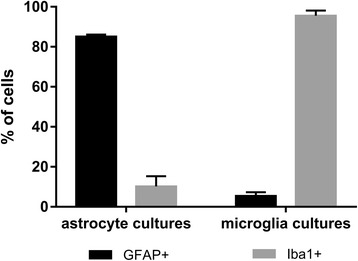
Fig. 1.

Purity of glial cell cultures used in the study as revealed by immunofluorescence. In astrocyte cultures, GFAP+ cells corresponding to astrocytes were dominant, while microglia cultures consisted prominently from Iba1+ cells corresponding to microglia. Data are shown as the mean ± standard error of the mean (SEM) of five independent samples
