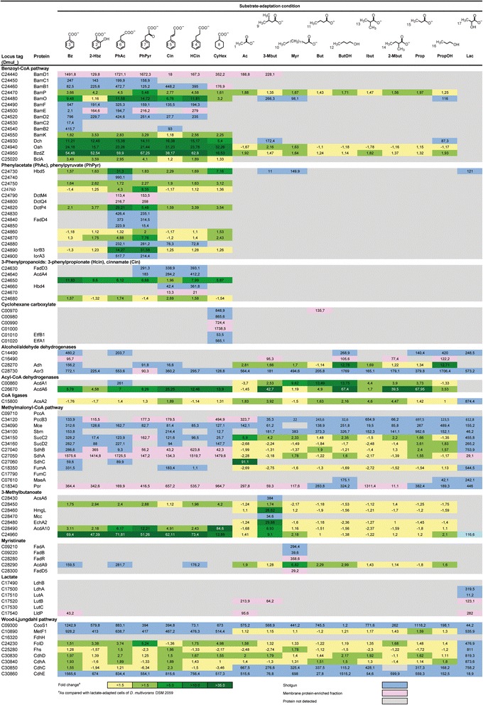
Fig. 3.

Subproteomes for the degradation of aromatic and aliphatic compounds across different substrate adaptation conditions in Desulfococcus multivorans. Compound names are as detailed in legend to Fig. 2. Identified proteins are ordered according to pathways/functional categories and ascending locus tags. Fold changes in protein abundance were determined by 2D DIGE using lactate-adapted cells as reference state. In case of multiple protein identifications, only one method is indicated hierarchically from 2D DIGE (green) via shotgun analysis (blue) down to membrane protein-enriched fraction (pink). Not detected proteins are indicated in grey. Enzyme abbreviations and functional predictions are according to Additional file 1: Table S1
