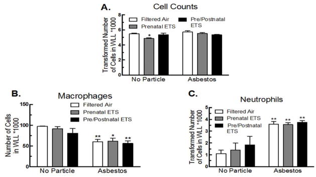
Figure 3. Inflammatory cell infiltration for FA, prenatal, and pre/postnatal ETS-exposed mice in WLL after 24 hr asbestos exposure.
(A) Total cell numbers, (B) macrophages, and (C) neutrophils. Data expressed as mean ± SEM. Asterisks indicate significance, ** at P < 0.01, * at P < 0.05 compared to FA, no particle. n = 6 mice per condition.

