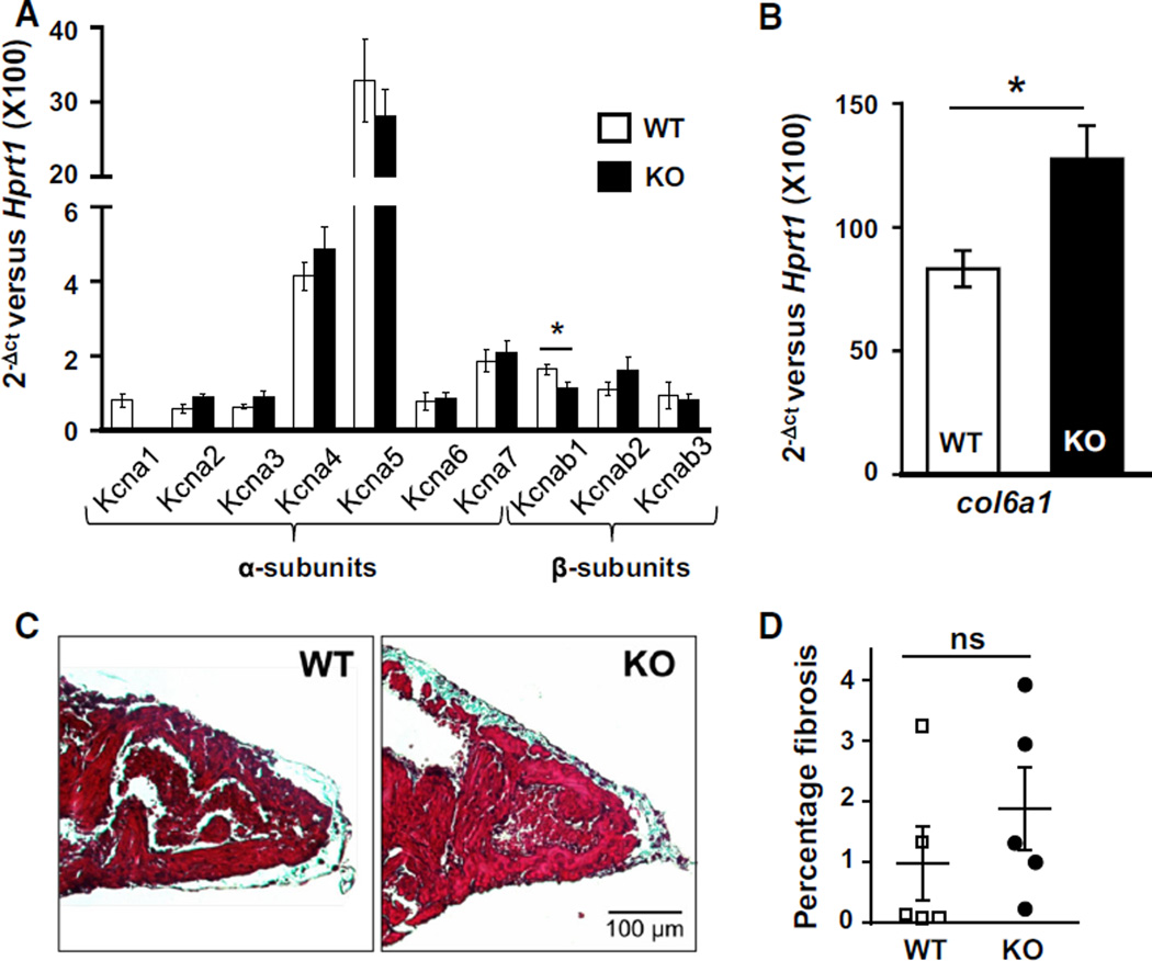
Fig. 2.
Kcna1-null mice exhibit minimal Kv1.x channel remodeling and atrial fibrosis. a Real-time PCR expression analysis of Kv1.x potassium channel α- and β-subunit genes in atria from KO and control WT mice (n = 5 mice per genotype). b Real-time PCR analysis of col6a1 mRNA levels (n = 5 mice per genotype). c Representative samples of atria from WT and KO animals stained with Masson’s trichrome to visualize fibrosis which appears bluish. Images were chosen out of sections from 5 WT mice and 5 KO mice. d Quantification of the percentage of atrial fibrosis between genotypes. *P < 0.05; ns not significant

