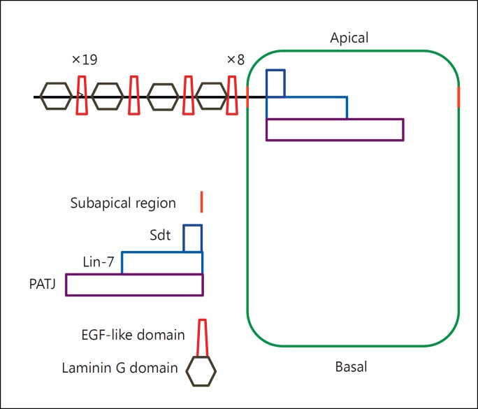
Fig. 2.
Schematic drawing of Crb in D. melanogaster, showing the position of the protein at the subapical region of the plasma membrane (marked in orange). The protein contains a large extracellular portion with numerous EGF-like domains (×19 and ×8 refer to repetitions of this repeat) and laminin G domains and a short intracellular portion that interacts with Sdt, Lin-7 and PATJ to form the Crumbs complex (after figs. 1 and 2B from Bulgakova and Knust, 2009).

