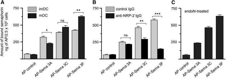
Figure 4. Sema3A, -3C, and -3F bind to the surface of human DCs.
(A) imDCs (gray bars) and mDCs (black bars) were incubated with recombinant AP-Sema3A, AP-Sema3C, AP-Sema3F, or lone AP (control), and binding of each ligand to the cell surface was determined by the amount of AP activity measured, as described in Materials and Methods. (B) mDCs were preincubated with preimmune goat IgG (control IgG; gray bars) or goat polyclonal anti-NRP-2 IgG (black bars) before exposure to Semas and subsequent measurement of bound AP activity. (C) mDCs were exposed to endoN to remove cell surface polySia before exposure to Semas and subsequent measurement of bound AP activity. In each experiment, each Sema (2.5 μg) was added to 5 × 10⁵ cells, and following the incubation, the samples were divided in half, and AP activity was measured. Data are the average from 3 sets of duplicate wells ± se from 1 experiment and are representative of results for 3 different experiments (*P < 0.05; **P < 0.01; ***P < 0.001; ns, P > 0.05).

