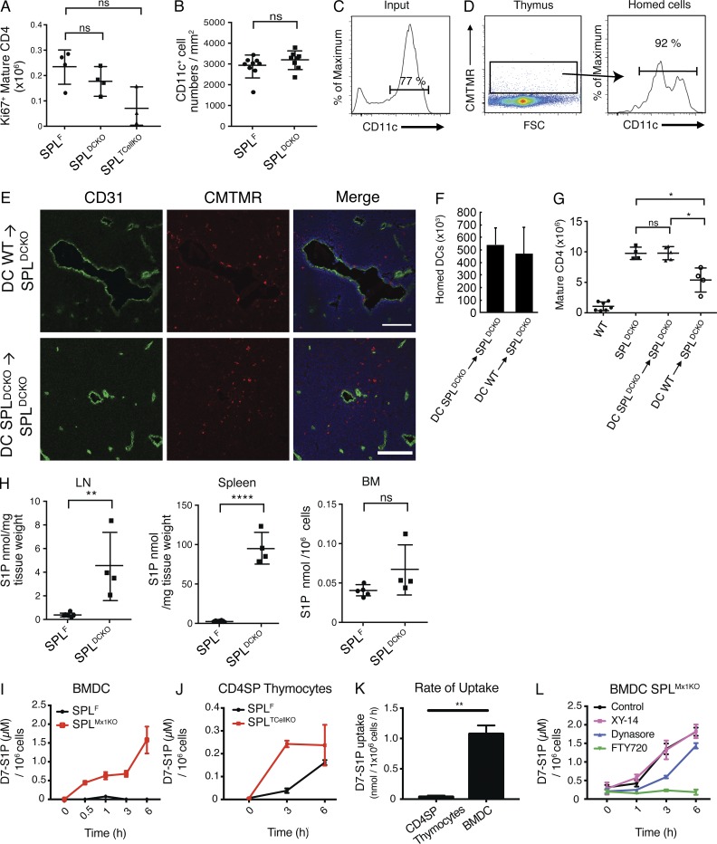
Figure 10.
WT DCs rescue the SPLDCKO thymic egress phenotype by uptake of S1P in a S1P1,3–5 receptor–dependent manner. (A) Absolute numbers of Ki67+ mature CD4SP T cells in SPLF, SPLDCKO, and SPLTcellKO mice. The results shown are representative of two independent experiments. (B) Number of medullary DCs per unit area. The graph is created from three thymuses per group. DCs were isolated from the spleens of either WT (DC WT) or SPLDCKO (DC SPLDCKO) donor mice as described in Materials and methods. Isolated DCs were administered intravenously into unirradiated SPLDCKO mice. (C) Flow cytometry of CD11c expression of the injected DCs (input). (D) 5 d later, thymuses were collected, and single-cell suspensions were analyzed. (Left) Thymocyte suspension showing homed CMTMR+ cells. FSC, forward side scatter. (Right) CD11c expression on gated CMTMR+ cells. (C and D) Numbers above the bracketed lines indicate the frequency of CD11c+ events. The results shown are representative of three independent experiments. (E) Immunofluorescence was performed on frozen thymic sections from SPLDCKO mice 24 h after injection using CD31 to stain the blood vessels and Hoechst (blue) to stain the nuclei. Bars, 100 µm. The results shown are representative of five thymuses analyzed. (F) Homing measured by flow cytometry for the presence CMTMR+ cells 5 d after injection. The graph represents a compilation of five thymuses per group. (G) 5 d after injection of DCs, SPLDCKO-recipient mice, as well as WT and SPLDCKO control mice, were euthanized, and mature (CD62LHiCD69Lo) CD4SP T cells were quantified by flow cytometry in whole thymuses. The graph represents a compilation of three independent experiments. (H) S1P levels in LN, spleen, and BM in SPLF and SPLDCKO mice. The graphs represent a compilation of two independent experiments. (I) Mature BMDCs from SPLF and SPLMx1KO were incubated with 1.5 µM D7-S1P, and their uptake was measure by LC/MS over time. The results shown are representative of two independent experiments. (J) CD4SP thymocytes isolated from SPLF and SPLTCellKO mice were incubated with D7-S1P, and their uptake was measured by LC/MS. The results shown are representative of two independent experiments. (K) Rate of D7-S1P uptake was calculated in SPL-deficient CD4SP thymocytes and mature BMDCs. The rate of change was calculated from two independent experiments. (L) Mature BMDCs were incubated for 24 h with either 10 µM XY-14, 5 µM FTY720, 80 µM of dynasore, or DMSO vehicle control before introducing 1.5 µM D7-S1P in the medium. Intracellular D7-S1P was quantified by LC/MS. The results shown are representative of two independent experiments. (A, B, and F–L) Mean values ± SD are shown. *, P < 0.05; **, P < 0.01; ****, P < 0.0001 for two-tailed unpaired Student’s t tests between the indicated groups.

