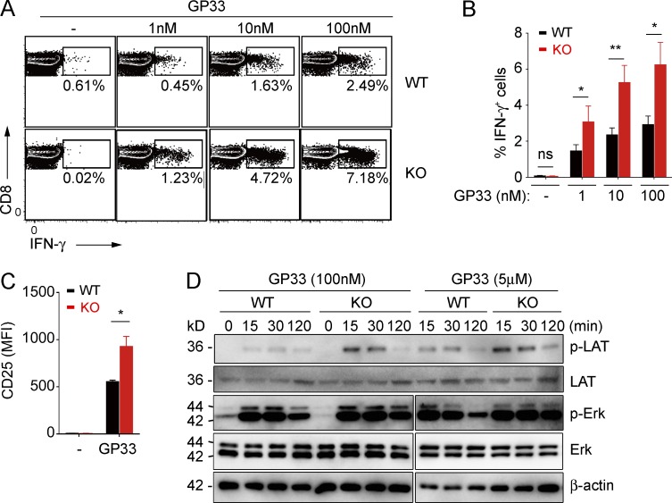
Figure 2.
Lack of TMEM16F causes increased T cell activation. (A–C) Flow cytometry analysis of intracellular IFN-γ (A and B), and surface CD25 (C) of CD8 T cells activated by GP33. Quantification of IFN-γ–producing cells is shown in (B). MFI, mean fluorescence intensity. Results are displayed as mean ± SEM. *, P < 0.05; **, P < 0.01; ns, not significant, using Student’s t test. B, n = 6; C, n = 3. (D) Phosphorylation of LAT and ERK induced by GP33 stimulation was detected by immunoblot of splenocytes from KO or WT mice. Data are representative of at least two experiments.

