Figure 7.
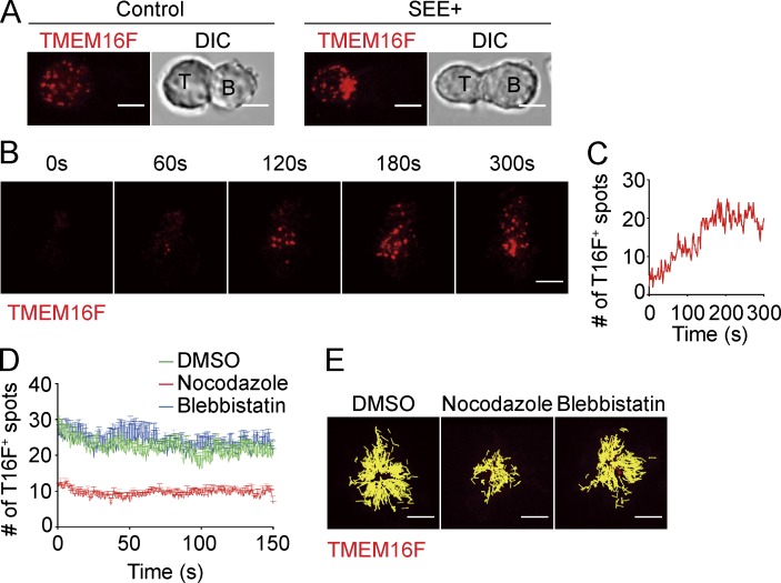
TMEM16F is recruited to the synapse and requires microtubules for transport. (A–E) Jurkat cells expressing TMEM16F-RFP were co-cultured with Raji cells (A) or stimulated on coverslips coated with αCD3 (B–E). (A) Confocal microscopy analysis for localization of TMEM16F in Jurkat T cells co-cultured with control (unpulsed) or SEE-pulsed Raji B cells (SEE+). 2-µm z stack of images is shown. DIC, differential interference contrast. (B and C) Dynamics of TMEM16F at the TCR activation site were imaged by TIRF microscopy. Number of TMEM16F-positive spots was quantified in C. (D and E) Dynamics of TMEM16F at the TCR activation site were imaged by TIRF microscopy in Jurkat cells pretreated with vehicle (DMSO), 1 µM nocodazole, or 1 µM blebbistatin. (D) Number of TMEM16F-positive spots was quantified. Time zero is the start of recording. n = 5 for each group. (E) Trajectories of TMEM16F-positive spots were tracked by ImageJ. Bars, 5 µm. Data are representative of three independent experiments.

