
Fig. 1.
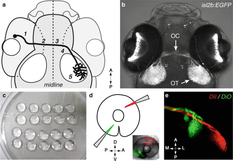
Methods for visualizing retinal axons. (a) Diagram of the retinal axon pathway. Retinal axons navigate to the optic nerve head (1), pass through the optic nerve and exit the eye (2), cross the midline at the chiasm (3), and grow dorsally along the optic tract (4) to reach the tectum (5). (b) Dorsal view of a Tg[isl2b:EGFP]zc7 transgenic embryo, in which EGFP is specifically expressed in all RGCs, allowing a direct visualization of retinal projections. Courtesy of A. Pittman. OC optic chiasm, OT optic tectum. (a, b) dorsal views, anterior up. Maximum intensity projection, confocal microscopy. (c–e) Focal injection of dyes in the retina allows visualization of retinal axons making topographic connections in the tectum. (c) Embryos are mounted laterally in low-melt agarose drops placed on a Petri dish lid. (d) DiI- (red) and DiO-(green) coated glass micropipettes are briefly inserted in a peripheral direction into the retina to label dorsonasal (DN in red) and ventrotemporal (VT in green) retinal neurons (method described in detail in Subheading 3.2). A anterior, P posterior, D dorsal, V ventral. (e) Dorsal view of the corresponding retinal axon projections in the brain target, the tectum. A anterior, P posterior, M medial, L lateral. Maximum intensity projection, confocal microscopy