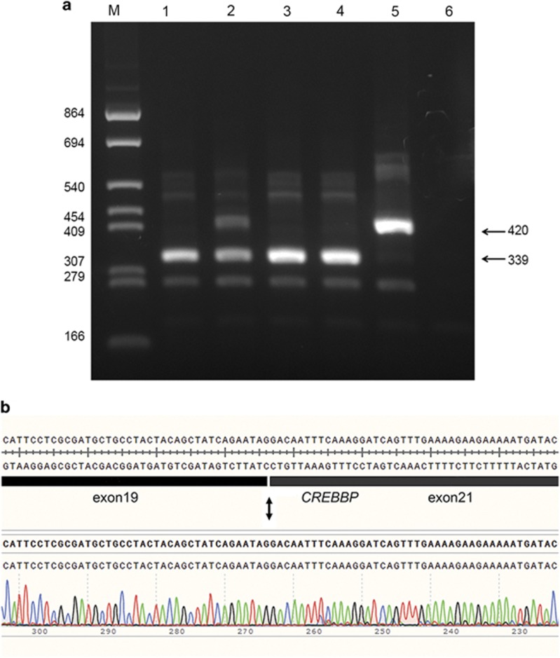
Figure 2.
Alternative splicing of CREBBP intron20 splice donor site mutations demonstrated by exon-trapping with the pCDNAGHE vector. (a) A 2.5% agarose gel showing the results of a RT-PCR with primers in CREBBP exon19 and GH1 exon5 on samples (1) pCDNAGHE137pat1, (2) pCDNAGHE137pat2, (3) pCDNAGHE137pat3, (4) pCDNAGHE137pat4, (5) pCDNAGHE137, (6) pCDNAGHE137inv. A 339 bp fragment is seen in all patients corresponding with a deletion of exon20, with only patient 2 showing additionally a wild-type band of 420 bp, which is also seen in the normal control. No bands can be seen for the inverted insert of pCDNAGHE137inv. (b) Sequence of the 339 bp fragment (patient 1) shows a CREBBP exon20 deletion.

