
Figure 3. In vitro.
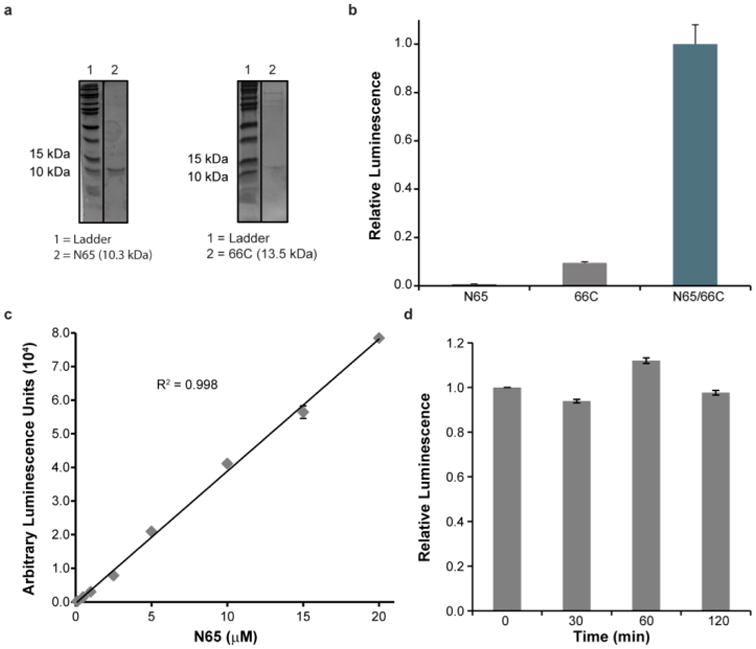
characterization of N65 and 66C Nluc fragments. (a) Coomassie stained gels of purified N65 and 66C. (b) Spontaneous reassembly of purified N65 (5 μM) and 66C (50 nM) fragments. Luminescence was measured 50 min after addition of substrate. (c) Luminescence of purified 66C (384 nM) in the presence of increasing concentrations of N65. (d) Luminescence of N65 (250 nM) and 66C (250 nM) fragments assessed at the indicated time after mixing by the addition of coelenterazine. Error bars represent the standard deviation of triplicate experiments.