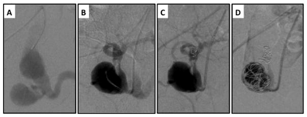
Figure 4.
Staged embolization of internal iliac artery prior to EVAR. A) Left iliac artery angiogram demonstrates aneurysms in both the common iliac artery and internal iliac artery. B) The hypogastric artery is catheterized and its distal bifurcation is visualized. C–D) Both the internal iliac artery bifurcation and aneurysm sac are embolized with Nitnol coils to prevent retrograde filling of the common iliac artery aneurysm sac following EVAR with planned distal fixation in the external iliac artery.

