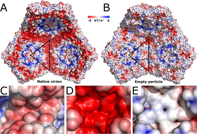
FIG 6.
Comparison of charge distribution inside the AiV-1 virion and empty particle. (A and B) Comparison of electrostatic potential distribution inside the native virion (A) and empty particle (B). Three pentamers of capsid protein protomers are displayed. The borders of a selected icosahedral asymmetric unit are highlighted with a black triangle. (C to E) Details of the electrostatic surface of the empty particle around 2-fold (C), 3-fold (D), and 5-fold (E) symmetry axes.

