Fig. 2.
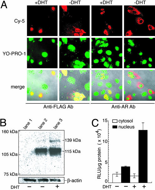
Characterization of the indicators in vitro.(A) The immunocytochemical images of COS-7 cells transiently transfected with pcRDn-NLS or pcDRc-AR. The COS-7 cells were cultured for 36 h, and then the cells were incubated for 2 h in the absence or presence of 1 μM DHT. (Top) The two expressed proteins were recognized by anti-AR and anti-FLAG antibodies, respectively, and stained with Cy-5-labeled secondary antibody. (Middle) The nuclei stained with YO-PRO-1; (Bottom) their merged images are shown with the transmission. (B) Western blot of protein extracts from COS-7 cells (lane 1) and from the cells cotransfected with pcRDn-NLS and pcDRc-AR in the absence (lane 2) or presence (lane 3) of 1 μM DHT. As a reference for the amounts of the electrophoresed proteins, β-actin was stained with its specific antibody. (C) Quantitative analysis of the Rluc activity for nuclear and cytoplasmic fractions. The cellular fractions were obtained from cotransfected cells with pcRDn-NLS and pcDRc-AR in the absence or presence of 1 μM DHT (n = 3).

