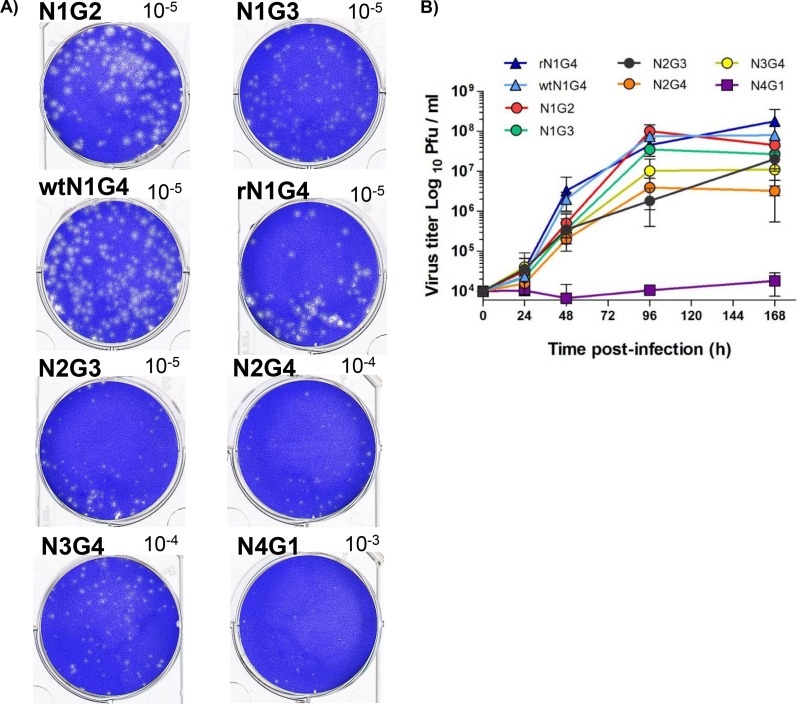
FIG 3.
(A) Plaque size differences among rIHNVs. EPC cells were infected at an MOI of 0.01 for 7 days, fixed, and stained with crystal violet. Corresponding well dilutions are given at the top corner of each well. (B) rIHNV kinetics of replication. Growth curves were determined for EPC cells in triplicate with an MOI of 0.01. Averages and standard deviations are shown.

