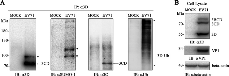
FIG 1.
EV71 3D is SUMOylated during infection. RD cells (1 × 10⁷) were infected with EV71 (MOI = 10) and harvested at 8 h postinfection before immunoprecipitation with an anti-3D antibody. IP and IB analyses were performed with the indicated antibodies in the presence of NEM. (A) Immunoblot detection by anti-SUMO-1, anti-Ub, anti-3C, and anti-3D antibodies during EV71 infection after immunoprecipitation by the anti-3D antibody. (B) Immunoblot analysis of cell lysis during infection. Anti-VP1, anti-3D, and anti-β-actin antibodies were used to detect the expression of VP1, 3D, and β-actin during infection. Lysis of RD cells without infection was set as a mock control. SUMO-1-modified 3D, ubiquitin-modified 3D, and 3CD are indicated by asterisks, brackets, and arrows, respectively.

