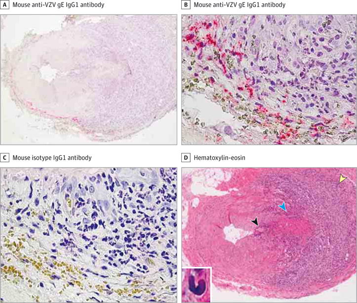
Figure 1. Identification of Giant Cell Arteritis Pathology in the Temporal Artery Adjacent to a Section Containing Varicella-Zoster Virus (VZV) Antigen in a Temporal Artery Signed Out as Pathologically Negative for Giant Cell Arteritis.

A-C, Immunohistochemical analysis using mouse anti-VZV gE IgG1 antibody revealed VZV antigen at the border of the adventitia and media of a temporal artery that was originally signed out as negative for giant cell arteritis (A and B, pink), but not when mouse isotype IgG1 antibody was substituted for mouse anti-VZV gE IgG1 antibody (C) on adjacent sections (original magnification ×100 [A] and ×600 [B and C]). D, Hematoxylin-eosin staining of a temporal artery section adjacent to that containing VZV antigen in A and B revealed giant cell arteritis pathology, with extensive inflammation in the adventitia (yellow arrowhead) and lesser inflammation in the media (blue arrowhead) and intima (black arrowhead) (original magnification ×100). Inset, Medial damage as well as numerous epithelioid cells were seen throughout the artery (hematoxylin-eosin, original magnification ×600).
