
Figure 2. Identification of Inflammatory Cells in a Section of the Temporal Artery Adjacent to Varicella-Zoster Virus (VZV) Antigen.
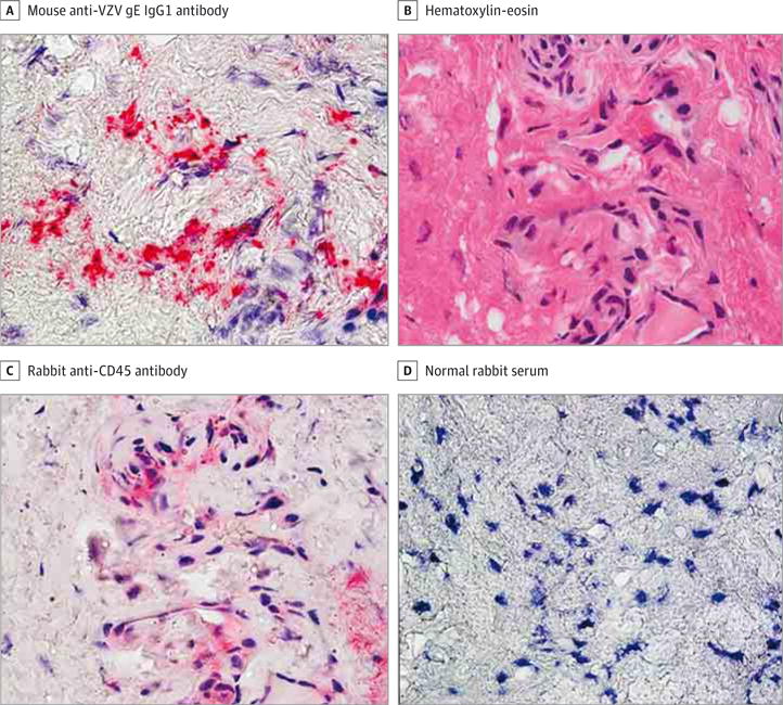
A, Immunohistochemical analysis using mouse anti-VZV gE IgG1 antibody revealed VZV antigen in the adventitia of a giant cell arteritis–negative temporal artery (bright pink) (original magnification ×600). B–D, Hematoxylin-eosin staining of a temporal artery section adjacent to that containing VZV antigen in A revealed inflammatory cells (B) expressing CD45 antigen with rabbit anti-CD45 antibody (C, pink) not detected when normal rabbit serum was substituted for rabbit anti-CD45 antibody (D) (original magnification ×600).