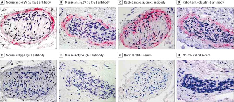
Figure 3. Colocalization of Varicella-Zoster Virus (VZV) Antigen and Claudin-1 in Cells in the Perineurium of Nerve Bundles in Temporal Arteries Pathologically Negative for Giant Cell Arteritis.

A, B, E, and F, Immunohistochemical analysis using mouse anti-VZV gE IgG1 antibody revealed VZV antigen predominantly in association with the perineurium of nerves in 2 representative temporal arteries (A and B, pink), but not in adjacent sections in which mouse isotype IgG1 antibody was substituted for mouse anti-VZV gE IgG1 antibody (E and F) (original magnification ×600). C, D, G, and H, Immunohistochemical analysis of sections adjacent to those containing VZV antigen with rabbit anti–claudin-1 antibody confirmed that the VZV antigen was located in association with perineurial cells expressing claudin-1 (C and D, pink), but not when normal rabbit serum was substituted for rabbit anti–claudin-1 antibody (G and H) (original magnification ×600).
