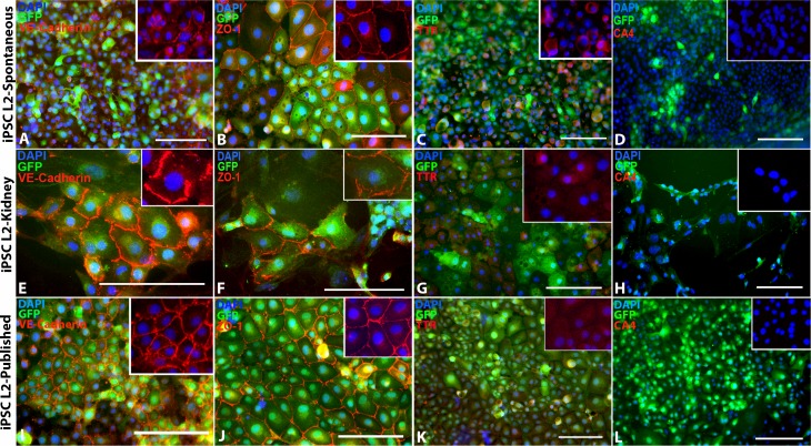
Figure 2.
Spontaneous iPSC-EC differentiation, kidney coculture iPSC-EC differentiation, and previously published iPSC- EC differentiation protocol. (A–D) Spontaneously differentiated iPSC-derived ECs express EC markers (A) VE-Cadherin and (B) ZO-1, a small amount of (C) TTR, but no (D) CA4. (E–H) iPSC-derived ECs differentiated in coculture with primary mouse kidney ECs also express (E) VE-Cadherin, (F) ZO-1, and (G) TTR, but not (H) CA4. (I–L) Likewise, iPSC-derived ECs differentiated using a previously published iPSC-EC differentiation protocol from Rufaihah et al.18 also express (I) VE-Cadherin, (J) ZO-1, and (K) TTR, but not (L) CA4. DAPI = blue. Scale bars: 100 μm.

