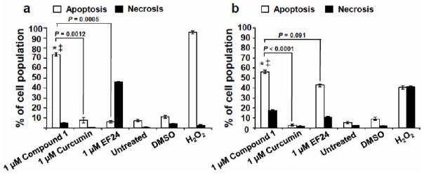
Fig. (3).
Compound 1 induced significant phosphatidylserine (PS) externalization on Jurkat and Nalm-6 leukemia cells after 24 h of treatment. PS exposure on the cell surface was monitored after treatment of two distinct lymphoma lines, Jurkat (a) and Nalm-6 (b). Apoptosis and necrosis were examined after incubation of the cell lines and co-staining with annexin V-FITC and PI and analyzed via flow cytometry assay. Each bar represents the average of three replicas with standard deviations. The total percentage of apoptotic cells is displayed as the sum of annexin V-FITC positive cells at early and late stages of apoptosis (white bars). PI-positive cells, but negative for annexin V-FITC, are considered undergoing necrosis (black bars). The following controls were included in these assays: treatment with the reference drugs curcumin and the EF-24 analogue (both at 1 μM); untreated cells as a negative control; DMSO as the solvent control; and H2O2-treated cells as a positive control for cytotoxicity. For both cell lines, a comparison of 1-treated cells with untreated (*) and DMSO (‡) controls, consistently resulted in P-values lower than 0.001. P-values comparing the percentage of apoptosis induced by 1 as compared with curcumin are annotated in the graphs. Data acquisition, processing and analysis were achieved by utilizing CXP software (Beckman Coulter).

