Figure 3.
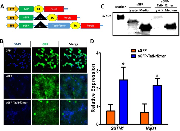
A secretable TatNrf2mer induces the expression of ARE genes. (A) Two lentiviral vectors delivering a secretable GFP (sGFP) or a sGFP fused to the TatNrf2mer by a furin cleavage site (FCS) were designed. Both constructs were cloned in-frame with the 2A-puroR sequence of the lentiviral vector to generate fusion proteins. Plasmids were packaged as lentiviral vector particles. (B) Distribution of GFP and sGFP-TatNrf2mer in stably transfected HEK293T cells. HEK293T cells were transduced with lentiviral vectors delivering either GFP, sGFP, or sGFP-TatNrf2mer and were selected in the presence of puromycin. The expression of GFP was evaluated by fluorescence microscopy. The sGFP and sGFP-TatNrf2mer had a different pattern of cellular distribution than the GFP expressing cells. DAPI staining was performed as a counter stain. (C) Stable cells were grown in low protein medium for 3 days. The conditioned media were collected and cells were lysed. The presence of GFP fused molecules was determined by Western blot using antibody to GFP. Although the sGFP-TatNrf2mer fused protein is detected in the lysate, the only band detected in the medium corresponds to cleaved GFP, thus suggesting the secretion and cleavage of the sGFP-TatNrf2mer protein. (D) The conditioned media from sGFP-TatNrf2mer increased the expression of two ARE genes in ARPE-19 cells. ARPE-19 cells were incubated with the 3-day conditioned media of either sGFP or sGFP-TatNrf2mer for 18 hours. Total RNA was isolated from these cells and a cDNA library was generated. The relative expression of the two ARE genes GSTM1 and NqO1 was measured by qRT-PCR using β-actin as a control transcript. Values are reported as average ± SD (n = 3 biological replicates).

