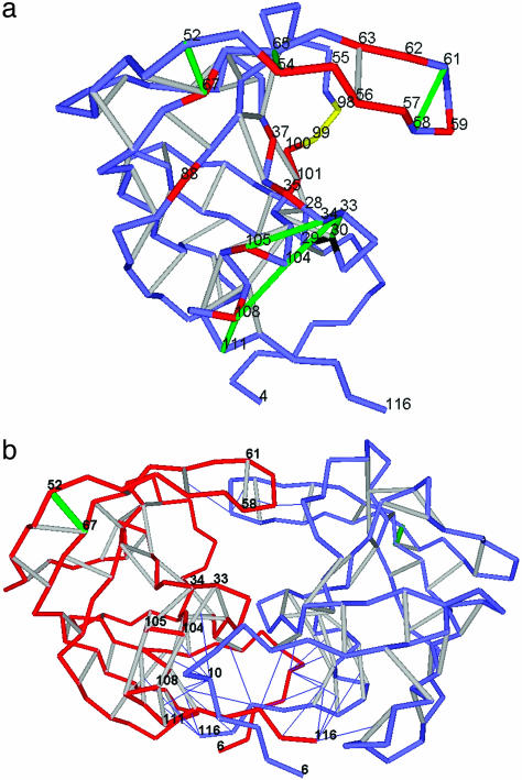
Fig. 2.
Packing defects in monomeric and dimeric (functional) FIV protease. (a) Dehydron distribution in FIV protease. The convention of Fig. 1 is followed. Mutation sites that affect substrate and inhibitor specificity (17) are marked in red if the substitution affects the wrapping of a dehydron and in yellow otherwise. (b) Dehydron distribution in the active dimeric FIV protease (PDB entry 3FIV). The intermolecular wrapping of dehydrons by L10 is highlighted. One monomeric chain (A) is depicted in red, and the other chain is depicted in blue. The color convention is the same as in a for dehydrons and the catalytic residue.

