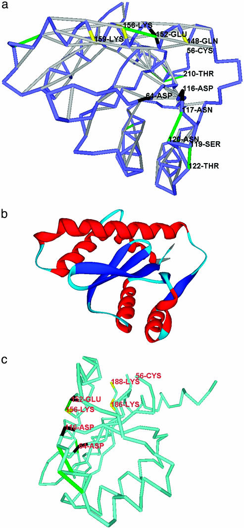
Fig. 3.
Packing defects in the stable fold of HIV-1 integrase. (a) Dehydron distribution for HIV-1 integrase (PDB entry 1B9F). The catalytic residues are marked in black, and the other active residues are marked in yellow. Dehydron (E152, K156), conventionally marked in green, constitutes a major epitope anchoring inhibitor drugs that dock along the major groove region parallel to the 146–164 helix. (b) Ribbon rendering of the HIV-1 integrase as a visual aid. Red, helices; blue, α-strands; light blue, turns and loopy regions. (c) The HIV-1 integrase positioned as in figure 4 of ref. 23, with the dehydrons (N117, N120) and (S119, T122) (a) defining the anchoring track for the viral DNA and the (E152, K156) defining the track for the host-cell DNA. The residue color convention is that of a, and the virtual-bond chain is displayed in lighter blue for visualization purposes.

