Fig. 2.

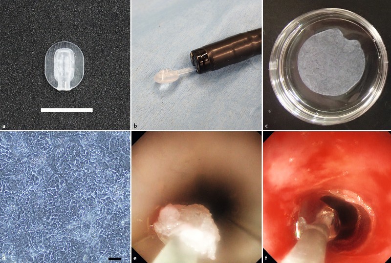
Transplantation of epidermal cell sheets (ECSs) using the newly developed device. a The head of the device was fabricated automatically by a 3 D printer. Scare bar indicates 10 mm. b The head docked with the catheter in front of the endoscope. c A tissue-engineered ECS (4.2 cm2) formed by primary cells isolated from pig skin fragments (2.5 cm2) after 2 weeks of culture. d H&E staining of epidermal cells in the ECSs, indicating a cobblestone-like pattern. Scale bar indicates 20 μm. (e, f) Actions of the developed device with two modes: transport and release. e The transport mode was set at suction with the ECS transported from the mouth onto the tears after endoscopic balloon dilation (EBD). f The release mode was set at air ejection. The release procedure enabled accurate placement of the ECS onto the artificial ulcer after EBD.
