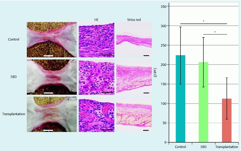
Fig. 4.

Histological findings. Macroscopic findings revealed severe strictures in control pigs, re-strictures in pigs that underwent endoscopic balloon dilation (EBD) alone, and the absence of re-strictures in pigs that underwent EBD followed by epidermal cell sheet (ECS) transplantation. White bars indicate 10 mm. H&E staining showed infiltration of inflammatory cells in all pigs (black bars indicate 50 μm). Sirius red staining revealed muscle atrophy and fibrosis in all pigs (black bars indicate 500 μm). The graph shows the numbers of inflammatory cells per high power field (HPF). *P < 0.01.
