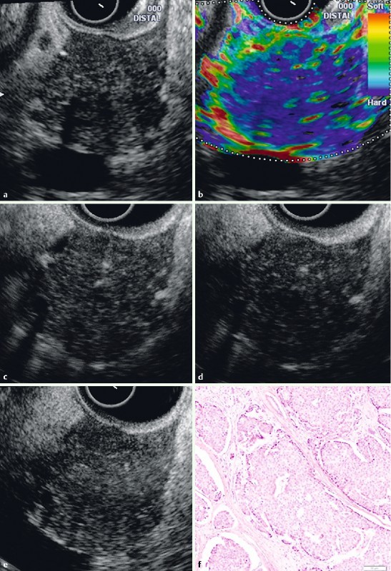
Fig. 1.

EUS B-mode image (a), EUS elastography, and contrast-enhanced EUS of a patient with an elastic score of 3 (patient number 2). Using EUS elastography (b), the lesion exhibits harder tissue (blue color) than the surrounding tissue. The borders of the lesion exhibit a red color (soft) in the elastography, which is correlated with minimal tumor invasion of the surrounding tissue via histology. The contrast-enhanced EUS study indicated a uniform increased enhancement at 1 minute (c), 3 minutes (d) and 5 minutes (e) in the contrast-enhanced harmonic mode. The histological results (f) indicated acinar cell carcinoma with an intraductal component. The fibrosis was minimal.
