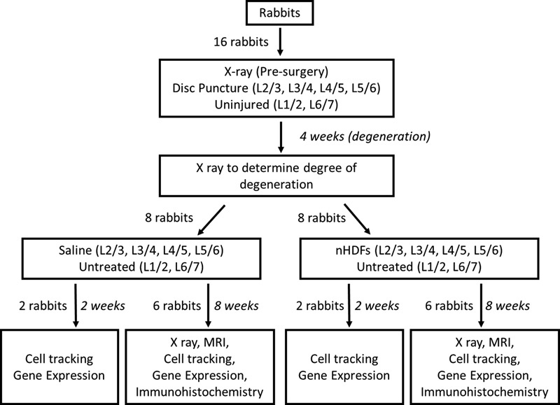
Fig. 1.

Study design. New Zealand white rabbits (n = 16) underwent surgery to induce disk degeneration by annular puncture on four disks (L2–L3, L3–L4, L4–L5, and L5–L6). Four weeks later, rabbits underwent a second surgery and were randomized to receive either neonatal human dermal fibroblasts (nHDFs) or saline treatment in the injured disks (L2–L3, L3–L4, L4–L5, and L5–L6). L1–L2 and L6–L7 intervertebral disks (IVDs) from each rabbit were left uninjured and untreated. Rabbits were euthanized at 2 weeks (n = 4, two per group) or 8 weeks (n = 12, six per group) post-treatment. X-ray images were obtained before the first surgery, 4 weeks after disk injury, and 8 weeks post-treatment. After euthanasia, rabbit spines underwent magnetic resonance imaging (MRI) and infrared imaging. Individual IVDs were further isolated to study changes in gene expression and collagen staining.
