Fig. 6.

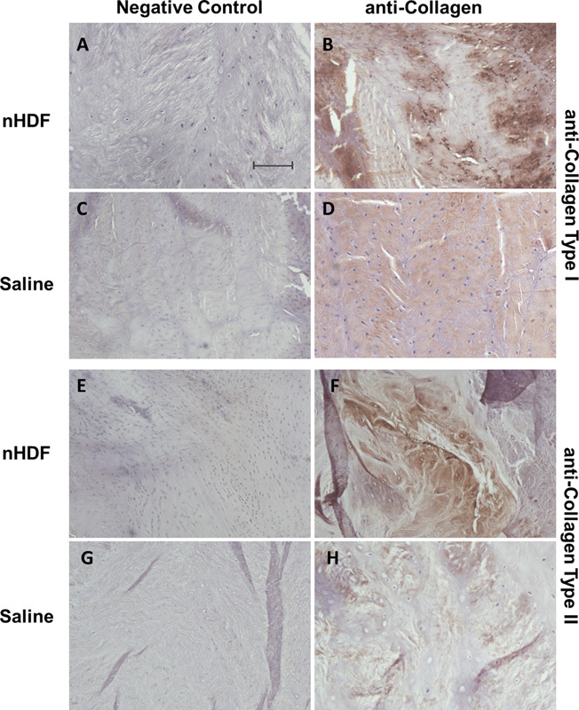
Representative immunohistochemical staining of collagen types I and II. Collagen type I staining of the annulus fibrosus regions in disks treated with (B) neonatal human dermal fibroblasts (nHDFs) or (D) saline and (A, C) their respective negative controls. Collagen type II staining of nucleus pulposus regions of disks treated with (F) nHDFs or (H) saline and (E, G) their respective negative controls. Negative controls had primary antibodies omitted. Note: Staining of both collagen types I and II is more intense in nHDF-treated disks compared with those treated with saline. Scale bar = 50 μm.
