
Fig. 1.
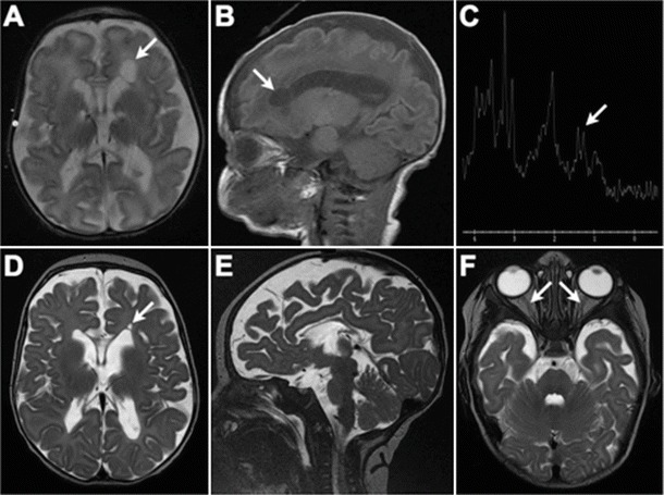
Cranial MRI appearance at 5 days (a, b) and 3 months of age (c–f). (a) and (b) Diffuse deficiency of cerebral white matter with enlarged fluid spaces and marked hypoplasia of the corpus callosum. Arrow indicates connatal (frontal lobe) cyst; a similar (smaller) right-sided cyst was present but is not well shown on this axial slice. (c) Lactate doublet is present on MR spectroscopy. (d) Evolution of changes in panels a and b. Myelination is markedly reduced for age. Basal ganglia, brainstem, and cerebellum have a relatively normal appearance. (f) Small optic nerves and optic chiasm (not shown)