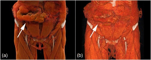
Fig. 8.

Non-enhanced 3-D CT images of traumatic herniation of small bowel through the anterior abdominal wall. CR (a) and VR (b) show the soft tissues of the abdominal wall and upper thigh along with herniating of the bowel loops (arrows). CR allows for a natural representation of anatomy
