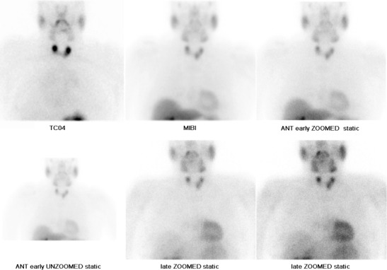
Fig. 10.

A nuclear medicine parathyroid scintigraphy study (image courtesy of Dr Cindy Leung, Barts and The London). The Technetium pertechnetate (99mTc-pertechnetate) image (upper row, far left) demonstrates localization of the thyroid gland. 99mTc-Sestamibi (MIBI) images show a focal area of significant increased tracer uptake inferior to the left thyroid lobe, which persists on early (centre and far right, upper row) and delayed imaging (centre and far right, lower row). Imaging characteristics are consistent with a parathyroid adenoma. Tracer uptake elsewhere is physiological
